Caring for your horse's feet
The saying ‘no foot, no horse’ is really important to understanding the essential need for regular good quality hoof care. It is important to make sure that your farrier is properly qualified and registered.
Under Section 16 of the Farriers (Registration) Act 1975 it is an offence for unregistered persons to carry out farriery. Farriery is defined in the Act as “any work in connection with the preparation of the foot of a horse the immediate reception of a shoe, the fitting by nailing or otherwise of a shoe to the foot, or the finishing off of such work to the foot”.
There is no definition of the word ‘shoe’ in the Farriers (Registration) Act 1975 but it is the Council’s view that the term refers to the purpose of a shoe and therefore includes conventional metal shoes and also non-metallic solutions, which may include, amongst others, glue-on plastic shoes or hoof wraps.
Illegal farriery is a criminal offence which can result in a fine of up to £1000, legal costs and a criminal record.
By using an unregistered person a horse owner may be risking the welfare of their animal, may invalidate any insurance if their horse is lamed or injured, and may be aiding and abetting criminal activity.
Over time your farrier will build up a relationship with your horse(s), and he or she will come to know your horse’s podiatry requirements.
Vets and farriers have good working relationships and here at Central Equine Vets we work closely with all of the farriers in the area. Find a registered farrier in your area here.
Nail bind and nail prick
A qualified farrier is an expert in his or her field, and the shoeing of horses will be very routine for him or her. When fitting shoes your farrier will most likely use nails to secure the shoe to the hoof (occasionally glue on shoes are used). Accurate placement of the nails is really important. This is to ensure that the shoe is held firmly on to the hoof, without causing damage to the sensitive laminae beneath the hard hoof wall.
Occasionally a nail can prick the sensitive laminae (nail prick). Most of the time the farrier will realise that this has happened straight away.
If the nail has been placed too close to the sensitive laminae, but hasn't actually contacted the laminae this is known as nail bind.
What are the clinical signs of nail bind and prick?
Lameness, of a varying a degree, occurring a few days after shoeing is the most obvious symptom. The hoof may feel warm to touch, and there may be an increased digital pulse present (compare with the hoof on the opposite limb). Application of hoof testers or tapping the hoof around the nail will produce a painful response.
Solar abscessation (pus in the foot)
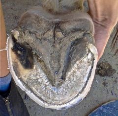
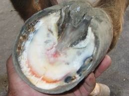
The most common cause of lameness is pus in the foot, so as an owner this is an important condition to know about! The lameness is usually confined to one leg and clinical signs will occur suddenly, often with the horse appearing sound one day and then becoming very lame the next day. The severity of the lameness is determined by the amount of pus produced and whether it has a means of escape. If there is no drainage then the pus builds up between the sensitive layers of the hoof and the hard hoof wall. As the hard hoof wall cannot expand this results in the pus putting pressure on the sensitive laminae. This results in pain and inflammation, which is really painful for the horse.
Figure 1: Typical appearance of a solar abscess
Causes of solar abscessation
Bacteria introduced into the foot by either a puncture wound or through a crack in the white line or hoof wall are the most common causes of solar abscessation.
What are the clinical signs of a solar abscess?
The following are the most common signs of solar abscessation in the horse:
Diagnosing a solar abscess
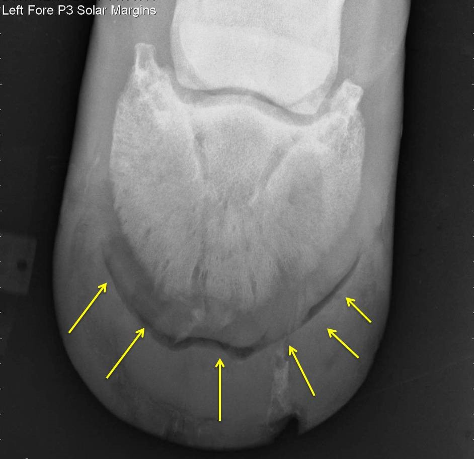
In most circumstances the clinical signs are enough to allow your vet to make a diagnosis of “pus in the foot”. The shoe should be removed to allow a full and complete examination of the foot. The presence of pus confirms the diagnosis, and of course helps to relieve the problem! However, occasionally an abscess, which releases pus, is not found straight away. At this point your vet will most likely advise twice daily warm, wet poulticing and review your horse in 2-3 days time to see if pus can be released then. Occasionally with very deep abscesses it may be necessary to poultice the foot for up to 7 days in an attempt to draw out the pus. Occasionally, nerve blocks may have to be performed to confirm the suspicion that the pain is definitely arising from the foot. If nerve blocks improve the lameness then the next step is to radiograph (X-ray) the foot. If an abscess is present this will appear as a gas shadow on the X-rays. Your vet will then be able to dig down on to the exact location of the abscess, and hopefully pus will be released!
Figure 2: X-ray of a solar abscess – note obvious gas shadow indicating the presence of pus
Treating a solar abscess
Once pus is released it is recommended that you use a wet poultice until you get two clean wet poultices. At this point you should then change to a dry poultice, and use these until you get two clear dry poultices. At this point, when the abscess has stopped discharging, the hole where the abscess has been released should be protected with a simple dry dressing to keep it clean until the defect begins to fill in with horn. The new horn, which forms over the site of the abscess, can be hardened more quickly by using swabs soaked in iodine (strength of weak tea) which are then wrung out and placed against the new horn filling the defect.
If an abscess is suspected, but not found at the time of the examination, then the suspected site of abscessation should be poulticed with a warm wet poultice and the horse re-examined in 48-72 hours.
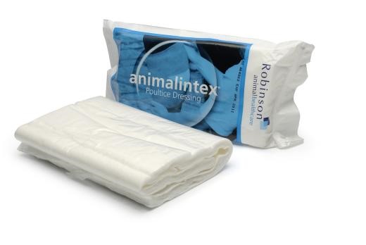
Figure 3: Poultice material – the mainstay of abscess treatment
How can I prevent a solar abscess from forming?
Your horse's feet should always be picked out (using your branded Central Equine Vets hoof pick!) and thoroughly cleaned before riding. Riding on uneven and stony ground should be avoided, particularly for thin-soled horses as this could increase the risk of solar puncturing. Regular attention from a competent farrier will ensure that your horses' feet are in optimal condition.
It is also vital to ensure that your horses are always fully vaccinated against tetanus, an invariably fatal infection which can gain access through hoof injuries.
Puncture wounds to the hoof
Like a human fingernail, the equine hoof is a tough structure, which helps protect the sensitive structures beneath. Most puncture wounds tend to affect the solar aspect of the foot. Depending on location and depth, puncture wounds to the solar aspect of the foot can sometimes prove to be very serious.
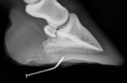
Your vet should always check out any solar puncture wound. Although, it may seem counter intuitive, any foreign body should be left in place (unless you feel that the foreign body is likely to be driven further into the foot when the horse weight bears. In which case it should be removed and the site of penetration marked). Leaving the foreign body in place helps the vet assess the likely severity of the puncture wound, and the foot can be radiographed (X-rayed) with the foreign body in place to help assess the structures, which may have been damaged.
Depending on the site of penetration structures, which may become damaged, are the laminae, pedal bone, coffin joint, navicular bursa and the deep digital flexor tendon. Danger areas, if penetrated, are the middle third of the frog and the back half of the foot, as the coffin joint, navicular bursa and deep digital flexor tendons can be involved. AS A RULE ALWAYS HAVE ANY SOLAR PENETRATION CHECKED OUT BY YOUR VET. REMEMBER TETANUS INFECTION CAN FOLLOW A SOLAR PENETRATION OR FOOT ABSCESS, SO ALWAYS VACCINATE YOUR HORSE AGAINST TETANUS.
Figure 4: Hoof penetration with a nail
Bruised Sole
The sole of the hoof is the insensitive protective undersurface bordered by the white line and the frog. The weight bearing areas of an unshod horse are the dorsal wall where it meets the sole of the foot and the frog. The sole of the hoof should not come into contact with the ground.
What causes bruised soles?
Beneath the sole lie the laminae. This is a sensitive and delicate blood filled tissue, which connects the sole to the pedal bone of the hoof and suspends the pedal bone within the hoof. Injury to the laminae causes bleeding between the sole and the pedal bone. This can form a bruise or haematoma, which is effectively a blood blister (like a brusied finger nail if you accidentally hit your finger with a hammer). This blood blister causes pressure within the non-expandable hoof wall, which results in pain, inflammation and lameness.
Solar bruises can be caused by your horse standing on a stone, excessive hard work on hard ground - especially if your horse is unshod or in ill fitting shoes.
What are the clinical signs of a bruised sole?
Contrary to popular belief the most obvious clinical sign of a bruised sole is lameness, and not an area of purple discolouration on the hoof. Any purple discolouration tends to happen much later on as the bruise “grows out”. The severity of lameness will vary according to the severity of the bruise and the horse’s pain threshold. The lameness usually develops as soon as the bruise develops. The lameness should be confined to the affected leg and pressure applied with hoof testers should demonstrate the area of the sole affected.
Figure 5: Bruised sole
How can I prevent solar bruises?
All horses' feet should always be picked and thoroughly cleaned out before exercise. Exercise on uneven and stony ground should be avoided, particularly for thin-soled horses or unshod horses. Specific food supplements containing biotin and methionine are also available which can help to promote healthy, strong hoof growth. Protective silicon hoof pads can also help.
Corns
Corns are bruises of the sole occurring at the seat of corn. This is the area of the sole, which lies between the bars and the wall at the back of the sole. However, unlike bruised soles where the lameness is evident almost immediately, corns can often develop over a long period of time.
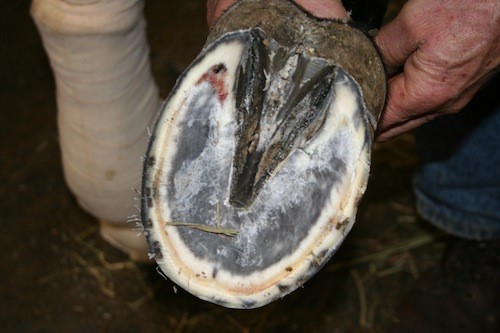
Figure 6: Corn (arrow points to the corn)
What causes corns?
Corns are caused when there is trauma to the seat of corn. The most common cause of a corn is shoes which are either too narrow or too tight and which put pressure on the seat of corn. Stones becoming trapped between the shoe and the seat of corn or shoes, which are left on for too long and begin to dig in, causing damage, are also potential causes of corns. Poor conformation such as low heels means that excessive weight is put on the heels and may cause trauma to the seat of corn, resulting in corns.
What are the clinical signs of corns?
Lameness is the most obvious clinical sign associated with corns. The severity of the lameness depends on how much damage has occurred. This lameness becomes more apparent if the horse is ridden on hard ground, in circles or lunged. Sometimes both front feet may be affected meaning it can be more difficult to diagnose corns. Pain can usually be elicited by using hoof testers to apply pressure over the affected seat of corn.
How do you prevent corns from forming?
A good farrier will ensure that your horse is shod properly, thus helping to prevent the formation of corns.
Thrush
Thrush is a condition affecting the frog region of the foot. Affected areas are usually soft and foul smelling. Although thrush can cause lameness, it is not commonly associated with the condition.
What causes thrush?
Thrush usually develops as a result of poor hoof hygiene, failure to clean out the feet regularly or if the horse is kept in damp and dirty conditions. If the hoof is continually damp and dirty it allows bacteria to invade, causing infection. The part of the hoof affected is the sulci (of which there are three) or grooves either side and in the frog. The bacteria involved is, Fusobacterium necrophorum. This is a particularly nasty anaerobic (air hating) bacteria, which eats away the layers of the frog and exposes the deeper more sensitive tissues. Horses with deep sulci are much more prone to developing thrush and horses with long toes and contracted heels will tend to develop deeper sulci, which may mean that infection is more likely.
What are the clinical signs of thrush?
Thrush produces a black, foul smelling, moist discharge in the affected sulcus of the frog. Affected areas can be painful when hoof testers are applied. Any foot can be affected.
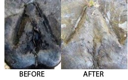
Figure 7: Typical thrush affecting the frog before and after treatment – note “eaten away” appearance of frog on the left
How can thrush be prevented?
Thrush is easily prevented by implementing good foot hygiene - daily cleaning of the stable and regular foot care and inspection, taking care to thoroughly pick out any debris which may have accumulated in the frog sulci. Regular attention from a farrier ensures the development of long heels is avoided and also to keep the frog healthy.
How to treat thrush
Prevention is always better than cure (see above). Areas affected by thrush should be trimmed away by the vet or farrier (the bacteria which causes thrush cannot live in an environment rich in oxygen – hence why it thrives in deep, dark unexposed areas of the foot). Following a trim of the frog topical antibacterial agents such as iodine sprays or antibiotic sprays can be used to kill further bacteria.
Hoof Cracks
Vertical cracks occurring in the hoof wall originate from either the bottom of the hoof or the coronary band. Cracks originating from the base of the hoof are called grass cracks while cracks originating from the coronary band are called sand cracks. They can be either partial cracks extending only part of the length of the hoof wall or complete cracks extending the entire length of the hoof wall. Cracks can be superficial, not affecting the sensitive laminae, or they can be deep where the laminae are affected.
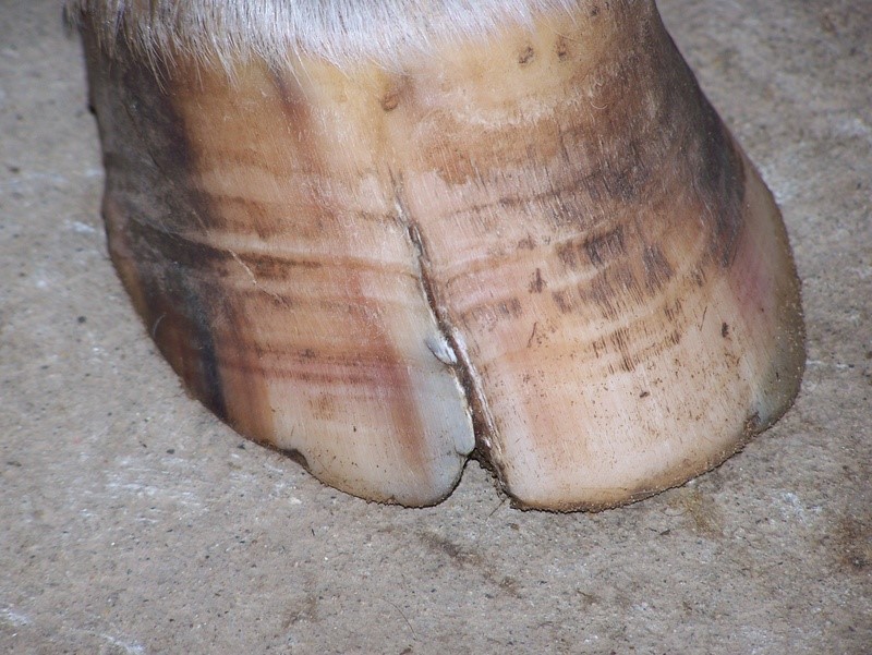
Figure 8: Typical grass crack
What causes hoof cracks?
Grass cracks are the most common type of hoof wall crack and are usually caused by overgrown hoof walls or unbalanced feet. Sand cracks, however, usually occur due to an injury to the coronary band or as a result of abnormal stress at the coronary band caused by unbalanced feet.
What are the signs of hoof cracks?
Hoof wall cracks are obvious, and depending on their length and depth some hoof cracks can also cause lameness. If a crack is superficial and does not affect the sensitive laminae then the horse may remain sound. If the crack is deep, and the laminae are involved or if there is movement associated with the crack then there will be a degree of lameness present.
How are hoof wall cracks treated?
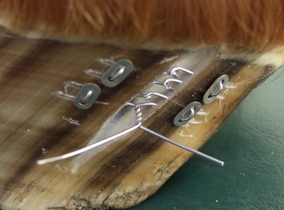
How they are treated really depends on the depth and location of the crack. Most of the time they will require to be stabilised. This is usually done by having the farrier fit a bar shoe. For mild cracks the farrier may “groove” the hoof horizontally to prevent the crack from extending further. Sometimes cracks need to be stabilised by using hoof staples, acrylic glues / fillers or a metal plate or bridge which is screwed into or glued on to the hoof to keep the crack stable while it grows out. Metal wire (a little like a shoe lace) can be fitted and tightened using a washer system to help stabilise some cracks too.
Figure 9: Repair of a quarter crack using a washer and wire system
How can I prevent hoof cracks from forming?
Regular attention from a competent farrier will ensure that your horse's feet are well balanced and in optimal condition. Specific feed supplements containing biotin and methionine are also available which can aid in hoof health, and help minimize the risk of some types of hoof crack from forming.