What is EGS?
Equine grass sickness (EGS) (equine dysautonomia) is a common and often fatal neurological disease affecting all equids. The condition is associated with degeneration of the autonomic, enteric (gastro intestinal) and somatic neurons (McGorum and Pirie 2009; Lyle and Pirie 2009). The disease is recognised throughout northern Europe and the United Kingdom, with the northeast of Scotland reported as having the highest incidence of grass sickness of 1-2% of the horse population per annum (McGorum and Pirie 2009). The disease occurs most commonly in the spring, following periods of cool and dry weather. Grass sickness is subdivided into three categories based on the severity and duration of clinical signs – acute, subacute and chronic. Acute EGS carries a hopeless prognosis usually resulting in death in less than 2 days. Subacute EGS also carries a hopeless prognosis with death usually occurring in 2-7 days. However, in cases of chronic EGS the duration of the disease is greater than 7 days (Doxey et al 1991) and, although the prognosis is guarded, some horses manage to fully recover (Milne and Wallis 1994; Milne 1997a,b,c; Lyle and Pirie 2009). This classification scheme suggests the duration of illness, but in actual fact more accurately reflects the severity of neuronal degeneration, particularly in the enteric (gastro intestinal) nervous system (Lyle and Pirie 2009). Some overlap and continuum also exists between the three categories (Pirie 2002).
The clinical signs observed are attributable to dysfunction of the autonomic (including the enteric) nervous system and commonly present as colic in the acute and subacute cases and weight loss or dysphagia in the chronic cases (Milne 1996; Proudman 2005; Lyle and Pirie 2009).
What causes EGS?
The cause of EGS remains elusive despite the condition initially being recognised over 100 years ago (Tocher et al 1923). The possible involvement of infectious and toxic agents including plants, fungi, insects, viruses, bacteria and their toxic compounds, mineral deficiencies and oxidative stress have all been hypothesised, however, none have been conclusively proven as causal agents of the condition (Pirie 2006; Lyle and Pirie 2009; McGorum and Pirie 2009). Recent research has lead to the current hypothesis that EGS is due to a toxicoinfection with Clostridium Botulinum types C and D (Hunter, Miller and Poxton 1999; Hunter and Poxton 2001; McCarthy et al 2004a; Nunn et al 2007; Lyle and Pirie 2009; McGorum and Pirie 2009). It is likely that a dietary trigger induces overgrowth of the resident gastrointestinal bacteria Clostridium Botulinum, with subsequent in vivo production of botulinum neurotoxins C, D, or both (Hunter, Miller and Poxton 1999; Hunter and Poxton 2001; McCarthy et al 2004a; Nunn et al 2007; Lyle and Pirie 2009; McGorum and Pirie 2009). However, further research is required to definitively prove this hypothesis (Pirie 2006; McGorum and Pirie 2009).
EGS was first reported in eastern Scotland in 1909 (Tocher 1923). Although recognised throughout the UK and most other northern European countries the highest incidence of EGS is in the northeast of Scotland, where it is reported to affect 1-2% of the horse population per annum (McGorum and Pirie 2009). Although the exact causal agent of EGS is currently unconfirmed, a number of important risk factors for the development of the disease are well recognised (table 1).
Table 1: Risk factors for the development of equine grass sickness.
|
Level of risk factor |
Risk factor and comments |
|
Horse related |
Age ; affects young horses aged between 2-7 years old Affects horses of good body condition Low serum concentrations of antibodies against surface antigens of C. botulinum type C and C. novyi type A and against C. botulinum type c neurotoxin complex toxoid Lack of recent contact with an affected horse |
|
Premises related |
Geographic location – horses in certain parts of the country and in certain fields and farms are often at greater risk Soil type – sand and loam soils have been associated with a higher risk of grass sickness compared to white chalk soils. Soil nitrogen content – high soil nitrogen content is positively associated with a higher frequency of cases Number of horses on premises – is positively associated with frequency of cases Previous occurrence – equine grass sickness is more likely to recur on premises with previous cases |
|
Management related |
Grazing or feeding of freshly cut grass is strongly associated with EGS Lack of co-grazing with ruminants increases the incidence of equine grass sickness, possibly through ruminants ingesting and removing the causal agent or due to co grazing reducing the stocking density of horses Recent movement (2-4 weeks), dietary change, stressful procedures – increase risk of equine grass sickness. This is thought to be due to the combined effects of an alteration in gastrointestinal flora and an immunological naivety to the aetiological agent Dietary changes likely reflect a change in the gastrointestinal flora, allowing for overgrowth of C. botulinum and subsequent neurotoxin production and absorption Pasture/ soil disturbance – building work causing soil disturbance may cause transfer of large numbers soil borne Clostridial species to the pasture surface Recent administration of ivermectin is associated with increased risk, but exact mode of action unknown, maybe a confounding factor Method of faeces removal – mechanical removal of faeces has been linked with an increased frequency of equine grass sickness in comparison to manual removal, presumably due to the potential for pasture and soil disturbance Frequent chain harrowing – associated with an increased frequency of cases due to pasture disturbance Grass cutting and supplementary feeding – associated with a reduction in the number of cases. Most likely due to reduced grazing intensity
|
|
Season/ climate related |
Cases of equine grass sickness peak in the spring and early summer and are usually associated with cool temperatures (7-11°C) and drier weather with irregular ground frosts in the two weeks prior to an outbreak. |
What are the signs and symptoms of grass sickness?
Clinical signs of grass sickness may be very variable depending on the form of the disease that the horse is suffering but can include patchy sweating, swallowing difficulties, recurrent impaction colic, dullness, lethargy, weight loss, depression and severe discomfort.
There are three categories of grass sickness:
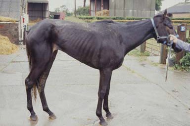
Picture 1: Horse with chronic EGS – note grey hound like appearance.

What are the chances of a horse surviving grass sickness?
Whether or not a horse survives grass sickness depends on the degree of damage suffered by the intestine and nervous system. Horses suffering from the more severe forms of grass sickness i.e. acute and sub-acute do not survive. Although there is no specific treatment, horses with chronic grass sickness may survive and go on to make a complete recovery with intensive long-term supportive care.
How is grass sickness diagnosed?
Diagnosing EGS is not straight forward as there is no definitive non-invasive antemortem diagnostic test for EGS. Most cases of EGS are diagnosed by recognising the associated clinical signs. However, the range of clinical signs that individuals can display is not consistent and varies according to the extent that the nervous system has been damaged. As such repeat examinations may be necessary before a diagnosis can be made. Sometimes it is necessary to take cases to surgery to discount other conditions, which may present similarly (such as a twisted gut or small intestinal blockage). Definitive diagnosis involves sampling a portion of small intestine (an ileal biopsy) during surgery, and examining it under a microscope. Grass sickness causes very specific damage that can only be identified microscopically.
One method that can be used as a diagnostic aid to assess the degree of nerve damage in cases of EGS is for the vet to administer a substance called phenylephrine into one of the horse's eyes. In a horse suffering grass sickness, the eye that received the drops will open slightly and the eyelashes will assume a different angle to that of the other eye. This test may be a helpful indicator along with other clinical signs, but it is not a definitive diagnostic aid because results can be variable.
References
1. Mcgorum, B.C. and Pirie, R.S. (2009). Grass Sickness. In Current Therapy in Equine Medicine 6, Eds.: N.E. Robinson, K.A. Sprayberry, Saunders Elsevier. pp 361-365.
2. Lyle, C and Pirie, R.S. (2009). Equine grass sickness. In Practice 31, 26-32.
3. Doxey, D.L., Gilmour, J.S. and Milne, E.M. (1991) A comparative study of normal equine populations and those with grass sickness (dysautonomia) in eastern Scotland. Equine vet. J 23, 365–369.
4. Milne, E.M. and Wallis, N. (1994) Nursing the chronic grass sickness patient. Equine vet. Educ 6, 217–219.
5. Milne, E.M. (1997a) Grass sickness In: Current Therapy in Equine Medicine 4, Ed: N.E.Robinson, W.B. Saunders Co., Philadelphia. pp 203–206.
6. Milne, E.M. (1997b) Grass sickness: an update. In Pract 19, 128–133.
7. Milne, E.M. (1997c) Clinical aspects and treatment of grass sickness in the UK In: Grass Sickness, Equine Motor Neuron Disease and Related Disorders: Proceedings of the 1st International Workshop, Eds: C.Hahn, V.Gerber, C.Herholz and I.G.Mayhew, Equine Veterinary Journal Ltd., Newmarket. pp 1–3.
8. Pirie, R.S. (2002) Grass sickness In: Manual of Equine Gastroenterology, Eds: T.Mair, T.Divers and N.Ducharme W.B. Saunders Co., London. pp 343–348.
9. Milne, E.M. (1996) Satellite Article. Clinical diagnosis and management of acute and subacute grass sickness. Equine Veterinary Education 8 (2), 71-73.
10. Proudman, C.J. (2005). Clinical Commentary. Equine grass sickness. Equine Veterinary Education 17 (1), 19-26.
11. Tocher, J.F., Tocher, J.W., Brown, W. et al (1923). Grass sickness investigation report. Vet. Rec. 3: 37-45, 75-89.
12. Hunter, L.C., Miller, J.K. and Poxton, I.R. (1999) The association of Clostridium botulinum type C with equine grass sickness: a toxicoinfection. Equine vet. J 31, 492–499.
13. Hunter, L.C. and Poxton, I.R. (2001) Systemic antibodies to Clostridium botulinum type C: do they protect horses from grass sickness (dysautonomia)? Equine vet. J 33, 547–553.
14. McCarthy, H.E., French, N.P., Edwards, G.B., Poxton, I.R., Kelly, D.F., Payne-Johnson, C.E., Miller, K. and Proudman, C.J. (2004a) Equine grass sickness is associated with low antibody levels to Clostridium botulinum: a matched case-control study. Equine vet. J 36, 123–129.
15. Nunn, F.G., Pirie, R.S., McGorum, B. Werney, U. and Poxton, I.R (2007). Preliminary study of mucosal IgA in the equine small intestine: specific IgA in cases of acute grass sickness and controls. Equine vet J. 39, 457-460.
16. Pirie, R.S. (2006). Grass Sickness. Clinical Techniques in Equine Practice 5, 30-36.