
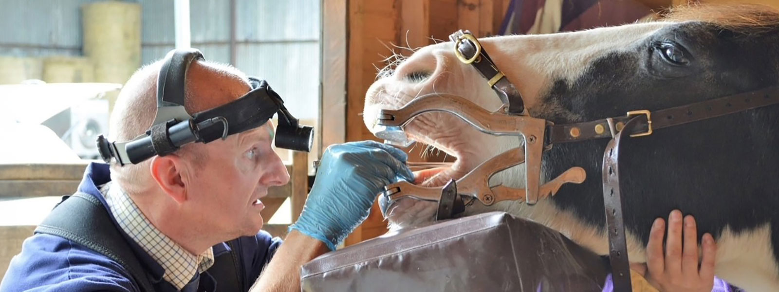
Lameness Examinations
Unfortunately, most horses suffer from a bout of lameness at some point in their lives. While the cause of lameness may be obvious, such as a foot abscess, corn or bruised sole, sometimes lameness can result from more complicated disorders.
Any lame horse requires a logical and thorough approach to accurately work out the cause of the lameness, so that an appropriate treatment plan can be devised with most investigations and procedures being completed remotely at the yard.
Initial examination
A general physical examination is usually performed first and after this, attention is directed towards assessing the limb(s), which may be affected. Following an examination of the limbs, the vet may carry out several initial investigations such as flexion tests and trotting the horse up to identify and localise where the pain is coming from. Occasionally it may be necessary to lunge the horse on both a soft and a hard surface to try to work out which limb(s) are lame.
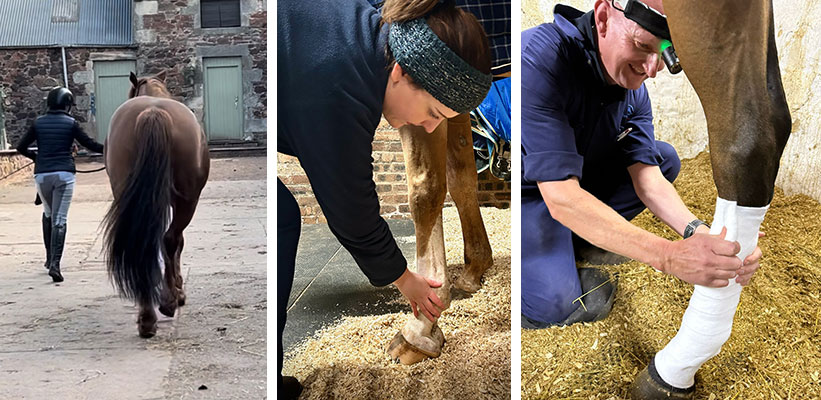
Horse being trotted up | Examination of a leg | Bandaging a wounded leg
Follow up investigations
Nerve blocks are the next logical step in the lameness investigation helping to isolate the area of pain more accurately. In turn this allows the vet to focus attention on the structures that are innervated by a particular nerve e.g. a particular joint, tendon, ligament etc.
Joint blocks can be performed however, you must wait for the nerve block to wear off to accurately assess joint blocks. If the problem area involves the joint e.g. as in a case of osteoarthritis, then the horse should become sound around 10 minutes or so post injection.
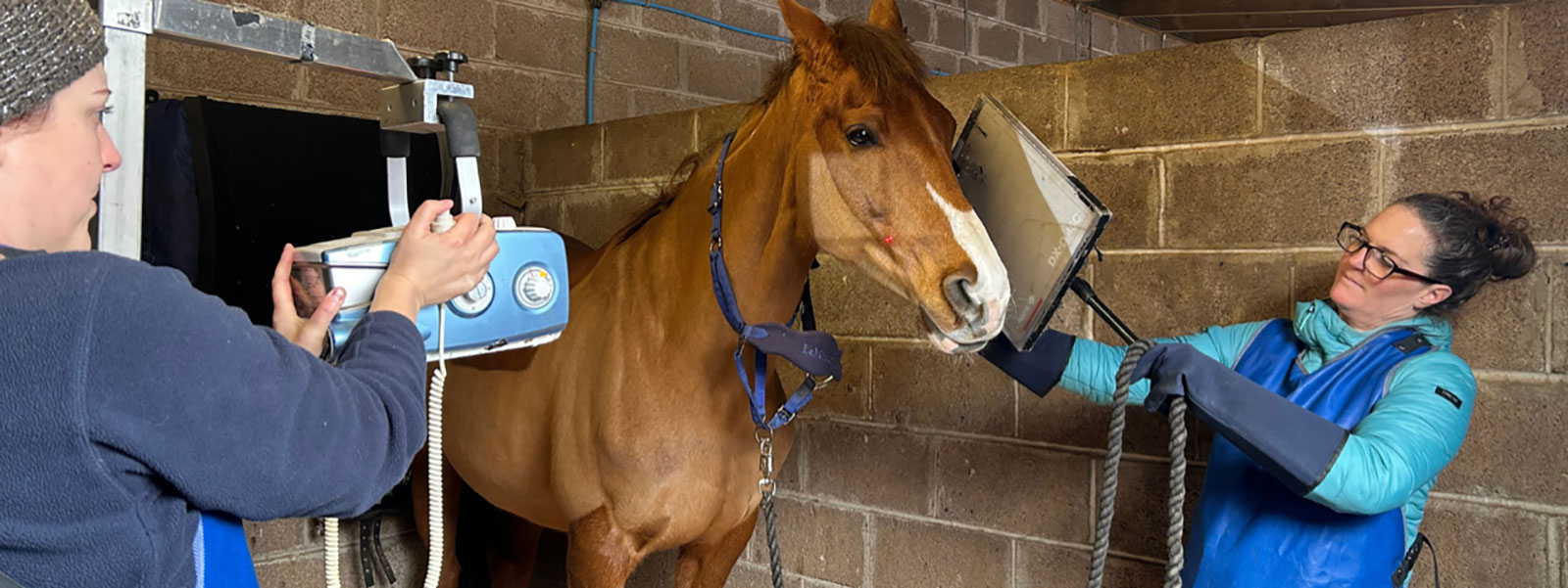
Radiographs can be completed once the seat of pain has been isolated and are particularly useful for assessing bone abnormalities e.g. cases of osteoarthritis, fractures etc.
Ultrasonography is also frequently used in lameness investigations and is most commonly used to assess potential damage to soft tissue structures such as ligaments and tendons.
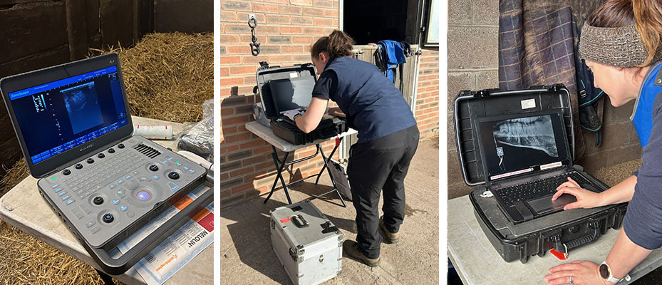
Portable ultrasound scanner | Portable x-ray machine being set up | Reviewing the images
In some cases, further, specialist investigation in a hospital may be required such as nuclear scintigraphy, (also known as a bone scan) or an MRI or CT which are more definitive advanced diagnostic imaging techniques.
Laminitis Investigation
Another common lameness can be as a result of horses suffering from laminitis. Laminitis is an extremely painful condition affecting the feet of horses. Laminitis is one of the most painful conditions that a horse or pony can suffer from. As such all cases of laminitis constitute a veterinary emergency.
Laminitis may be caused by:
- Endocrine (hormonal) conditions (Equine Cushing’s Disease / PPID and Equine Metabolic Syndrome)
- Massive carbohydrate overload
- Toxic insults (e.g. post colic surgery, colitis, retained placenta)
- Support limb laminitis
Generally, treatment of acute laminitis relies on box rest, anti-inflammatory and pain-relieving drugs, such as phenylbutazone (bute), and adequate support of the feet either through specially designed frog supports or deep bedding.
The veterinary surgeon will recommend that radiographs are taken as part of the laminitis investigation providing valuable information about any rotation and sinking of the pedal bone, which may have occurred. This, alongside a customised management plan will allow the veterinary surgeon to manage the active laminitis, to try and prevent worsening of symptoms and get to the bottom of why the laminitis has occurred.
As well as radiographs, once the horse is recovering from the acute (sore) phase, veterinary investigation will be aimed at trying to identifying the underlying cause of the laminitis which could be due to several reasons such as cushings disease, equine metabolic syndrome or both.
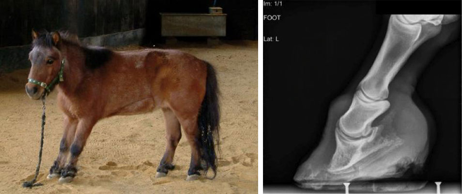
Typical laminitic stance in pony | Radiograph of the foot showing pedal bone rotation & descent due to laminitis
Central Equine Vets class laminitis as a veterinary emergency and recommend that should you have any concerns about your horse, you contact us straight away.
Should you have any concerns regarding lameness in your horse, please get in touch to organise an initial lameness examination to put your mind at rest.